Case Study: 62-Year-Old Female with Calf Pain and Swelling
Clinical Presentation
A 62-year-old female presented with two weeks of left calf tenderness and swelling (Figs. 1 and 2). She reported acute medial calf pain that began suddenly while walking and worsened with dorsiflexion.

Fig. 1
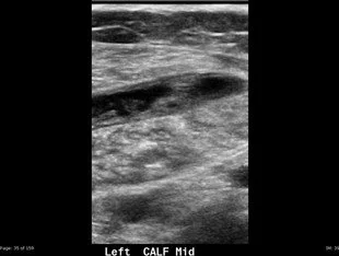
Fig. 2
Diagnostic Steps
Ultrasound Findings:
Linear collection of fluid and echogenic material separating the superficial gastrocnemius muscle (G in Figs. 3 and 4) from the deeper soleus muscle (S in Figs. 3 and 4).
Comparison to the normal tendon anatomy (arrows in Figs. 5 and 6) shows significant disruption at the expected location of the plantaris tendon.
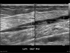
Fig. 3

Fig. 4
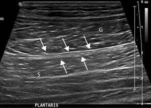
Fig. 5
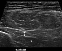
Fig. 6
Differential Diagnosis
Possible conditions include:
Gastrocnemius muscle tear
Soleus muscle tear
Dissecting or ruptured Baker cyst
Final Diagnosis - Plantaris Tendon Rupture
Key Teaching Points
Anatomy and Incidence:
The plantaris tendon lies between the medial gastrocnemius and soleus muscles.
Approximately 10% of individuals lack one or both plantaris tendons.
Mechanism of Injury:
Tears typically occur during activities involving sudden acceleration or deceleration, such as running or jumping.
Frequently referred to as "tennis leg."
Can occur in isolation or alongside other injuries, such as gastrocnemius, soleus, or ACL tears.
Clinical Features:
Patients often describe a "pop" during activity, such as stepping off a curb or lunging.
Acute medial calf pain, swelling, and exacerbation of pain with dorsiflexion are common.
Can mimic deep venous thrombosis or muscle tear/sprain
US Findings:
The normal plantaris tendon appears as a slender, echogenic structure with a distinct fibrillar pattern, running between the bellies of the gastrocnemius and soleus muscles.
In the event of a complete tear, the plantaris muscle retracts proximally, leaving the torn tendon tract filled with fluid or hematoma. This results in a linear, tubular, heterogeneous fluid collection located between the gastrocnemius muscle anteriorly and the soleus muscle posteriorly.
Differential Imaging Features:
Tears of the gastrocnemius or soleus muscles present as hypoechoic, heterogeneous areas that disrupt the normal pennate sonographic architecture of the muscle.
In this case, the abnormality is specifically situated at the interface between the gastrocnemius and soleus muscles.
A dissected or ruptured Baker's cyst, by contrast, typically appears as a tear-shaped, heterogeneous fluid collection located superficially to the gastrocnemius muscle.
Management:
Treatment is primarily conservative.
Active stretching exercises should be avoided, as they may worsen the tear.
Reference
Scoutt LM, Hamper UM, Angtuaco TL, Scoutt LM, Hamper UM, Angtuaco TL, eds. Case 145. In: Ultrasound. Oxford University Press; 2016:0. doi:10.1093/med/9780199988105.003.0145